
Quantitative Cell Cycle Analysis Based on an Endogenous All Biology Diagrams We demonstrate how to mitigate the effects of cell cycle heterogeneity in scRNA-seq data by calculating cell cycle phase scores based on canonical markers, and regressing these out of the data during pre-processing. Keywords: cell cycle, labeled nucleosides, BrdU, EdU, markers of cell cycle phases, DNA labeling, time lapse microscopy 1. Introduction A common task of many research teams is the analysis of cell cycle progression through the distinct cell cycle phases. Various methods can be used for this purpose. These antibodies to top cell senescence markers are critical for studying cell cycle arrest in research fields like development, aging, and cancer.

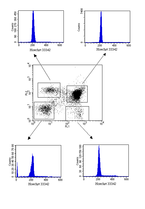
According to the complex changes in cell structure and biosynthesis, the cell cycle is divided into four phases: gap 1 (G1), DNA synthesis (S), gap 2 (G2), and mitosis (M). Determining which cell cycle phases a cell is in is critical to the research of cancer development and pharmacy for targeting cell cycle. Do Propidium Iodide staining and so Dlow cytometry. This will tell you where your cells are being arrested in cell cycle. Now so Cyclin and CDKs of that specific phase for confirmation.

Basic Methods of Cell Cycle Analysis Biology Diagrams
Determining which cell cycle phases a cell is in is critical to the research of cancer development and pharmacy for targeting cell cycle. However, current detection methods have the following problems: (1) they are complicated and time consuming to perform, and (2) they cannot detect the cell cycle on a large scale.
Here we review classical approaches that rely on cell fixation to characterise the cell-cycle status and its regulatory enzymes, and we describe the more recent development of cell-cycle markers based on genetically encoded fusions of fluorescent proteins with characteristic cell-cycle features, and of fluorescent biosensor technology to probe

cycle markers and biosensors Biology Diagrams
Some of the G2/M checkpoint (CHK2 etc) markers can also be useful but again there are some differences between cell culture/cell lines and tissue derived cells.
